症例
Allen CE. Langerhans-Cell Histiocytosis. N Engl J Med. 2018 Aug 30;379(9):856-868. doi: 10.1056/NEJMra1607548. PMID: 30157397. こちら。
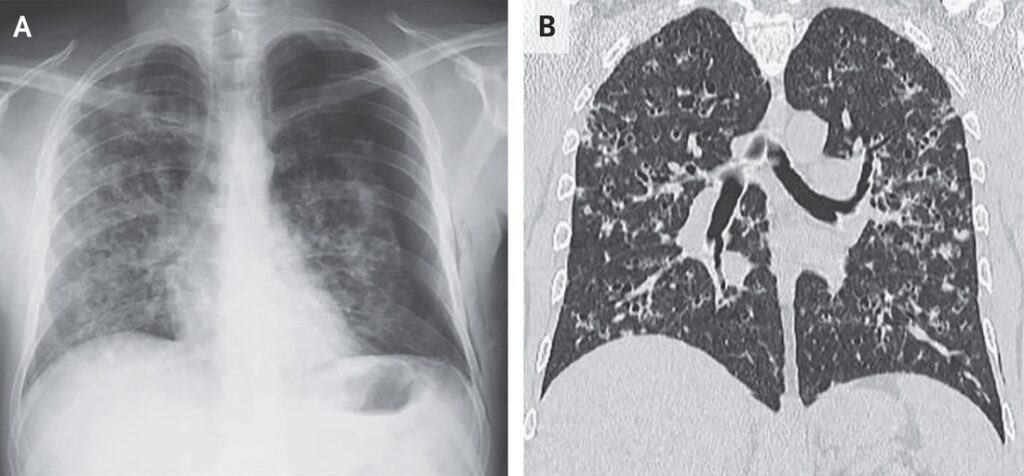
A 40-year-old man with a history of smoking presented to the emergency department with a 2-week history of cough, dyspnea, night sweats, and pleuritic chest pain on the left side. Physical examination was notable for decreased breath sounds over the left lung fields. A chest radiograph showed a large pneumothorax on the left side and interstitial infiltrates in both lungs (Panel A). The pneumothorax was treated with chest-tube thoracostomy. Subsequent computed tomography of the chest showed multiple cysts and nodules, predominantly in the upper and middle lung fields, with sparing of the costophrenic angles (Panel B). A transbronchial lung biopsy was performed. Histopathological tests showed a lymphocytic lung infiltrate with interalveolar septal thickening, eosinophils, and large cells with foamy cytoplasm and large nuclei (Panel C). Immunohistochemical staining was positive for S-100 protein, CD1a, placental acid phosphatase, and langerin. A diagnosis of pulmonary Langerhans-cell histiocytosis was made. Further testing revealed no evidence of systemic histiocytosis. BRAF testing was not done. The patient was advised to stop smoking, and a tapering dose of prednisone was prescribed. At the 6-month follow-up, the patient had ceased smoking; he was still taking low-dose prednisone, and his symptoms had abated.
40歳男性。喫煙歴がある。2週間にわたる咳嗽,呼吸困難,寝汗,左胸部痛の既往を訴えて救急外来を受診した.身体所見で左肺野の呼吸音が減弱している.胸部X線写真では、左肺の気胸、両肺に間質性浸潤が認められた(パネルA)。気胸に対して、胸腔チューブを挿入し、人工呼吸管理がなされた。その後、胸部CTで、上・中肺野を中心に多発性の嚢胞と結節が認められた(パネルB)。経気管支肺生検が行われた。
病理組織学的検査では,リンパ球性肺浸潤があり、肺胞間隔の肥厚,好酸球浸潤を認める。また泡沫状の細胞質と大型核を有する細胞を認める(パネルC).
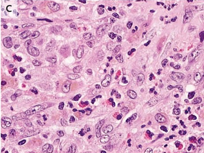
免疫染色では,この大型細胞はS-100蛋白,CD1a,胎盤酸性フォスファターゼ (胎盤性ALP),ランゲリンに陽性である.
最終的に肺ランゲルハンス細胞組織球症と診断された.全身性組織球症病変は認められなかった.BRAF検査は行われなかった。患者には禁煙指導,プレドニゾン投与がされた.6 ヵ月後のフォローアップでは,患者は禁煙が継続され,少量のプレドニゾン服用にて,症状は軽減した.
ランゲルハンス細胞組織球症の総説。
Charles M Harmon. Langerhans Cell Histiocytosis: A Clinicopathologic Review and Molecular Pathogenetic Update. Arch Pathol Lab Med. 2015 Oct;139(10):1211-4. doi: 10.5858/arpa.2015-0199-RA. PMID: 26414464. こちら。
病理
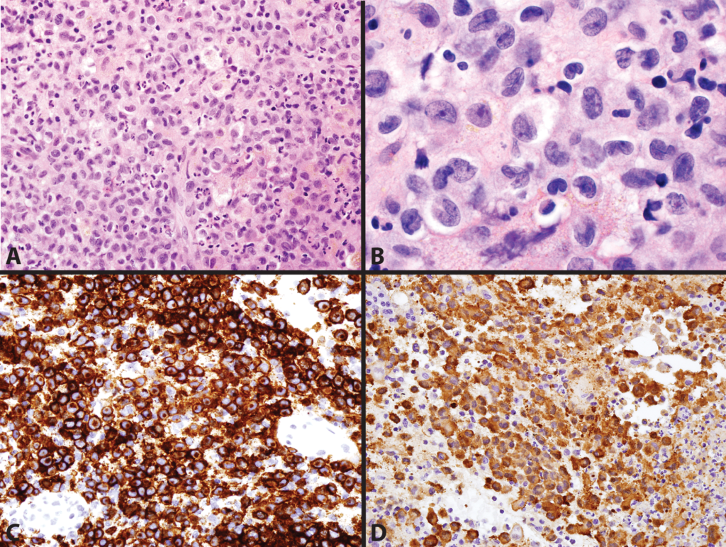
Morphologic and immunophenotypic features of Langerhans cell histiocytosis (LCH). A, Hematoxylin-eosin–stained section of a vulvar biopsy shows an infiltrate composed predominantly of large cells with abundant eosinophilic cytoplasm and irregular nuclei. Neutrophils, lymphocytes, and foamy histiocytes are present in the background. B, Prominent nuclear folds and grooves, fine chromatin, and small nucleoli are appreciated at higher magnification. C and D, The LCH cells are immunoreactive for antibodies directed against CD1a (C) and S100 protein (D) (hematoxylin-eosin, original magnifications ×400 [A] and ×1000 [B]; original magnification ×400 [C and D]).
ランゲルハンス細胞組織球症(LCH)の形態学的および免疫染色像。A) 外陰部生検のヘマトキシリン・エオジン染色切片では,豊富な好酸性細胞質と不規則な核を有する大型細胞から主になる浸潤を示す.好中球,リンパ球,泡沫性組織球が背景に存在する。B) 顕著な核のひだや溝、微細なクロマチン、小さな核小体が高倍率で観察される。CおよびD、LCH細胞は, C)CD1a および D)S100蛋白 の免疫染色で陽性(HE染色、×400[A]、×1000[B]、×400[C、D])。
LCHとBRAF V600Eバリアントについて こちら
組織球症は非常に様々な予後を示す原因不明の稀な疾患である。BRAF遺伝子の機能獲得変異であるV600E変異は、LCHの57%、Erdheim-Chester病(ECD)の54%に認められるが、他の組織球症にはみられない。メラノーマにおいては、変異BRAFの阻害剤(ベムラフェニブ vemurafenib)による標的療法で生存率は向上する。BRAF遺伝子のV600E変異のある治療抵抗性の、多臓器型ECD 3例、皮膚とリンパ節病変のあるLCH 2例に対するvemurafenib治療について報告する。臨床的、生物学的(CRP値)、組織学的(皮膚生検)、形態学的(PET、CT、MRI)に患者を評価した。全例において、vemurafenib治療により、迅速に明らかな臨床的および生物学的効果が得られ、治療開始1か月後のPET/CT/MRI検査により腫瘍の縮小がみられた。1例目では、治療開始後1~4か月の間でPET所見はどんどん改善した。依然として疾患活動性は残っているが、4か月の経過観察の間、治療効果は持続した。新たに承認されたBRAF阻害剤であるvemurafenibによる治療は、重症で治療抵抗性のBRAF V600E変異のある組織球症に対して、特に生命にかかわる病状では考慮されるべきである。
森本 哲 組織球症の病態解明と治療の進歩 こちら
難治性の LCH に対して, BRAF 阻害剤を導入する試みが報告されている.BRAF V600E 変異陽性で, vinblastine,cladribine に不応の高リスクの乳児 LCH に対し vemurafenib単剤療法を行ったところ,速やかに著効を得たという報告がある.最新の報告では,BRAF V600E 変異陽性難治性LCH に対し vemurafenib 単剤療法(20mg/kg 経口投与)を2 か月以上行い,16 例(高リスク 14 例,低リスク 2 例)中1 例(硬化性胆管炎合併例)を除き治療開始後 2 週間内に効果が得られた。
Sébastien Héritier. Vemurafenib Use in an Infant for High-Risk Langerhans Cell Histiocytosis. JAMA Oncol. 2015 Sep;1(6):836-8. doi: 10.1001/jamaoncol.2015.0736. PMID: 26180941 こちら
写真は走水神社から振り返って見た海 こちら


コメント